What is Mohs Surgery?
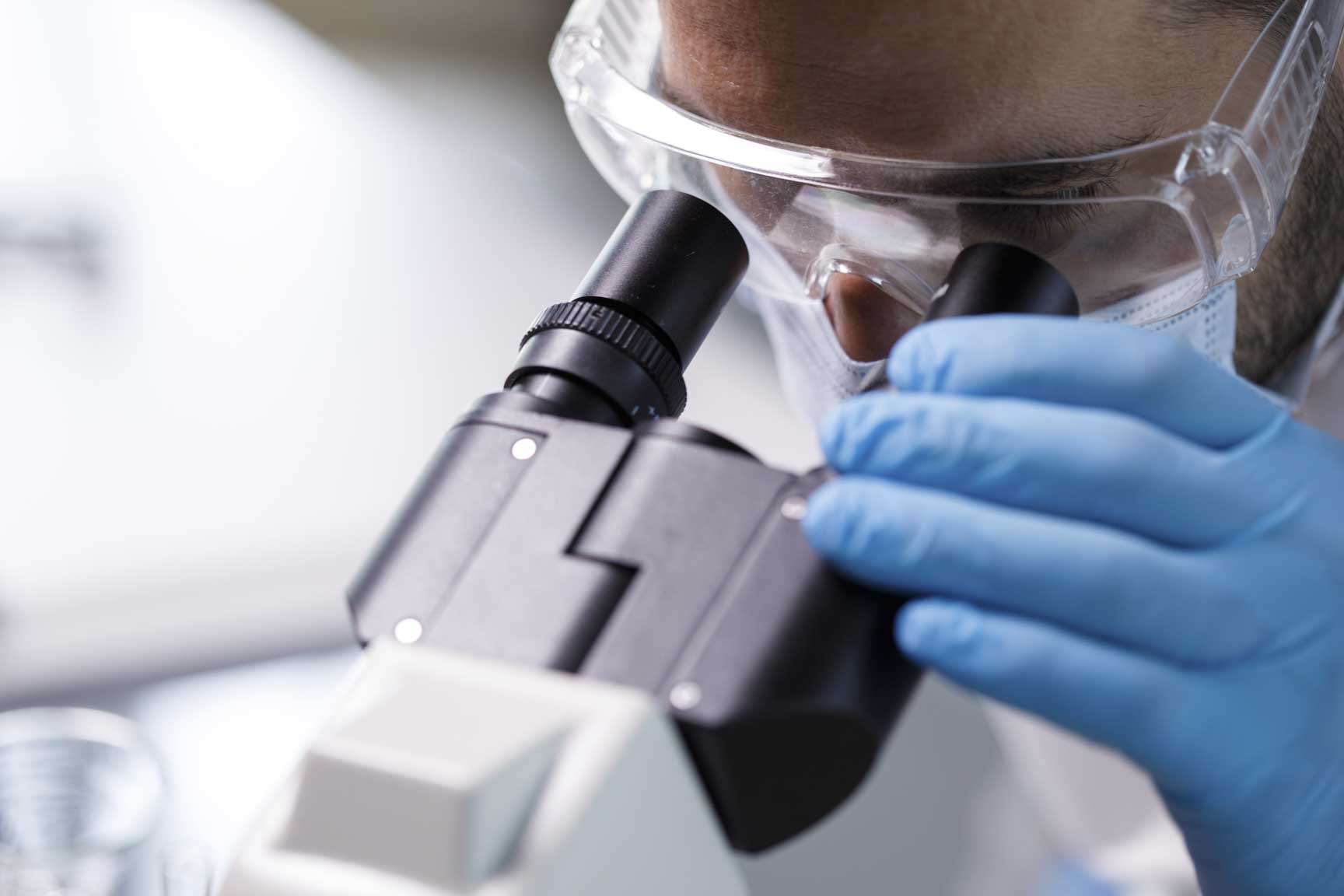
Mohs Micrographic Surgery is an advanced surgical technique for the treatment of certain skin cancers. It is most commonly used to treat squamous cell carcinoma and basal cell carcinoma.
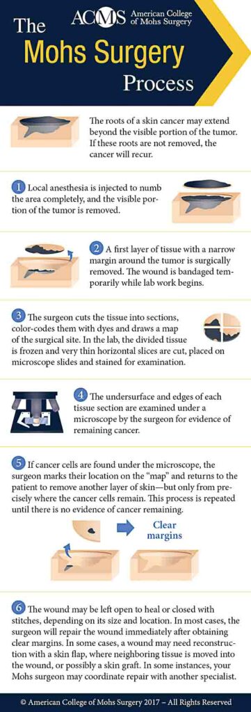
All cancer cells in the skin may not be visible to the naked eye. Cancer cells form roots below the surface of the skin and if these roots are not removed, cancer will recur. It is common for skin cancers to appear completely removed after the initial biopsy.
Pioneered by Dr. Frederick Mohs, Mohs micrographic surgery has been embraced by an increasing number of surgeons for an ever-widening variety of skin cancers. Mohs is the only skin cancer procedure that spares the greatest amount of healthy tissue while also most completely removing cancer cells.
The reason for the technique’s success is its simple elegance. During a Mohs surgery, microscopic examination of all excised tissue occurs during, rather than after the surgery. The procedure entails removing one thin layer of tissue at a time; as each layer is removed, its margins are studied under a microscope for the presence of cancer cells. If the margins are cancer-free, the surgery is ended. If not, more tissue is removed from the margin where the cancer cells were found, and the procedure is repeated until all the margins of the final tissue sample examined are clear of cancer. In this way, Mohs surgery eliminates the guesswork in skin cancer removal, producing the best therapeutic and cosmetic results.
Advantages of Mohs Surgery
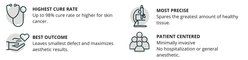
- Up to 98% cure rate or higher for skin cancer
- Spares the greatest amount of healthy tissue
- Minimally invasive. No hospitalization or general anesthetic
- Leaves smallest defect and maximizes aesthetic results
History
Mohs surgery is named after its founder and originator, Frederic E. Mohs, MD (1910-2002), who pioneered the procedure during his time at the University of Wisconsin in Madison. In June 1936, Dr. Mohs treated his first patient, who had squamous cell cancer of the lower lip.
The present-day American College of Mohs Surgery was founded in 1967 by Dr. Mohs as the American College of Chemosurgery. Over the years, the Mohs surgical process was refined and in 1988, membership changed the name to American College of Mohs Micrographic Surgery and Cutaneous Oncology. This was shortened on the College’s 40th anniversary in 2007 to American College of Mohs Surgery (ACMS)
Effectiveness
Mohs surgery has the highest cure rate of any type of skin cancer treatment; 97-99% for primary tumors and 94% for recurrent tumors. The cure rates of other methods are lower: 89.9 % for standard excision; 81-96% for destruction, and 91% for radiation. This high cure rate leads to fewer recurrences than with any other method.
Indications
Mohs surgery is the standard of care when a tumor is recurrent, has ill-defined margins, is in a critical cosmetic or functional location, or is large (> 2 cm) or aggressive.
Procedure
- Local anesthesia is injected to numb the area completely, and the visible portion of the tumor is removed.
- A first layer of tissue with a narrow margin around the tumor is surgically removed. The wound is bandaged temporarily while lab work begins.
- The surgeon cuts the tissue into sections, color-codes them with dyes and draws a "map" of the surgical site. In the lab, the divided tissue is frozen and very thin horizontal slices are cut, placed on microscope slides and stained for examination.
- The undersurface and edges of each tissue section are examined under a microscope by the surgeon for evidence of remaining cancer.
- If cancer cells are found under the microscope, the surgeon marks their location on the “map" and returns to the patient to remove another layer of skin-but only from precisely where the cancer cells remain. This process is repeated until there is no evidence of cancer remaining.
- The wound may be left open to heal or closed with stitches, depending on its size and location. In most cases, the surgeon will repair the wound immediately after obtaining clear margins. In some cases, a wound may need reconstruction with a skin flap, where neighboring tissue is moved into the wound, or possibly a skin graft. In some instances, your Mohs surgeon may coordinate repair with another specialist.
Reconstruction
After removal of the skin cancer in its entirety, the resulting defect is reconstructed to minimize scar appearance and preserve function. Dr. Campanelli and Dr. Dhanaraj are trained in aesthetic and functional repair and will repair the wound after skin cancer removal in the office. A wound may be left to heal on its own, or it may be repaired with side-to-side stitches, a flap, or a skin graft from another site on the body. In some cases, we may work with another specialist to repair your wound.
Cost Effectiveness
Mohs surgeons often perform removal, pathology and reconstruction functions under local anesthesia in a single outpatient office visit, eliminating the need for expensive operating room time, general anesthesia, multiple hospital visits and multiple specialists. An extremely low recurrence rate ensures patients do not incur costs for additional procedures.
The Mohs Surgeon
While any board certified dermatologist may perform Mohs surgery, only ACMS members have undergone rigorous fellowship training. Chosen through an extremely competitive review and selection process, fellows are required to complete an intensive 1 or 2-year post-residency ACMS fellowship training program.
Dr. Campanelli and Dr. Dhanaraj are fellowship trained surgeons in programs recognized by the ACMS, and have extensive experience in Mohs surgery, pathology, and reconstructive surgery.